
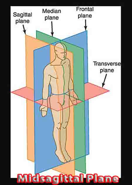
The Route of the Human Midsagittal Plane
Generally, the midsagittal plane depicts the human in anatomical position with arms and legs slightly apart, as shown here.
Left and right midsagittal sections are referred to as left and right sides of the subject, not to the point of view of the person looking at, for example, a computed tomography image. Sagittal planes (also called parasagittal planes) can divide an organism into unequal vertical sections along its longitudinal axis, whereas the midsagittal plane provides two equally-sized halves.
An organ or point of reference close to the midline is described as medial. A lateral point is one that is farther from the mid-line.
Head
Starting at the top of the skull, the midsagittal or median plane runs through the frontal, parietal, occipital and nasal bones, the maxilla and the mandible. The following image shows the median section of the human skull.
In the soft tissues of the head, the median plane separates the brain into its two hemispheres, together with the near-equal separation of nasal cavity, tongue, pharynx, and larynx, the latter .
The entire vertebrae – cervical, thoracic, and lumbar – are vertically divided into two halves, through the spinal cord. The image shows the median division between thoracic and lumbar vertebrae.
Thorax
The soft and hard tissues of the thorax are vertically divided into two sections with one lung and one set of ribs on either side. Due to the non-symmetrical divisions within the human anatomy, the contents of each midsagittal section are never exactly the same.
The aortic arch, for example, features more in the left-hand section than in the right, and the trachea, placed very slightly to the right-hand side, will be most prominent in the right-hand section. An anterior (taken from the front) X-ray clearly shows the heart in its asymmetrical position, while the 3D picture next to it shows this in more detail. When divided at the midsagittal plane, the ventricles are primarily placed within the left midsagittal section, and the atria to the right, bearing in mind left and right sides correspond to the subject’s orientation and are opposite to viewer perception.
Abdomen
From the above picture, one can also see that the position of ‘symmetrical’ organs like the kidneys does not always provide a mirror image. This is particularly true in the abdomen. Additionally, the abdomen contains the pancreas, stomach, appendix, liver and gall bladder, all of which are singular and placed either completely or partially within a single midsagittal section.
Above, the large liver is positioned within the right half of the body and the left-hand kidney is decidedly higher than that of the right-hand side. The bladder and bones of the pelvis are centrally placed and feature in relative symmetry on both sides of a midsagittal image.
Urogenital systems in males and females are, on the whole, symmetrical. A single midsagittal section of the female reproductive system features one ovary; that of the male will contain one testis. Bladder, uterus, penis and urethra are divided through the middle.
Abnormal Midsagittal Sections in Humans
Many anatomical abnormalities can lead to unusual results on medical images. Spinal deformities such as scoliosis prevent a midsagittal section from running neatly through the middle of each vertebra.
Genetic defects leading to the lack of an organ, such as renal agenesis or the absence of one or both kidneys during fetal development, will lead to an abnormal imagery result. Situs inversus, where major organs appear opposite to the normal human anatomy, can affect the position of the heart (dextrocardia).
Situs inversus totalis is a rare condition where every thoracic and abdominal organ is found in a mirrored position. This does not lead to medical complications, except where pre-surgery imaging is omitted. The image below shows the reversed position of the heart in an individual with situs inversus.
Dextrocardia
Craniomaxillofacial (CMF) deformities are another cause of irregularities between left and right midsagittal sections. Usually the result of facial traumas or genetic irregularities, facial dissymmetry can cause further problems such as respiratory or visual difficulties.
In cases of reconstructive craniomaxillofacial surgery, specialists try to return trauma victims to the premorbid (pre-trauma) midsagittal plane; in cases of genetic deformities, to a normal anatomical structure.
Supernumerary anatomy describes situations where the body contains an extra part, organ or part of an organ. These additions can either be symmetrical, such as in the case of Myrtle Corbin (pictured below) or asymmetrical.
Usually, supernumerary anatomy is the result of disruption during embryonic development through genetic mutations. Mutations in the fetus are sometimes caused by drugs such as thalidomide when taken by the mother during pregnancy.