
Muscle Cell Definition
A muscle cell, or myocyte, is a specialized animal cell that can shorten its length by using a series of motor proteins. In addition to several other associated proteins, actin and myosin form filaments that slide past each other to contract muscle cells. They are called sarcomeres, and they run end-to-end within a larger fiber called a myofibril.
Several nuclei are pressed against the cell membrane in a single muscle cell. Compared to other types of cells, muscle cells are long, and many of them connect to form the long fibers found in muscle tissue.
Structure of a Muscle Cell
The myofibrils that make up a muscle cell are compact bundles. Sarcomeres are bundled together and attached end-to-end to form myofibrils. Within and around these myofibril bundles, the sarcoplasmic reticulum extends from the endoplasmic reticulum. Signals from nerve cells activate the sarcoplasmic reticulum (SR), which concentrates a chemical that allows muscle cells to contract.
As a result of the nerve signals traveling through the transverse tubules, the SR is activated. To maintain a constant flow of ATP, mitochondria are densely packed within muscle cells. Cells are covered with a specialized membrane called a sarcolemma. Sarcolemma has special openings that allow nerve impulses to pass into transverse tubules.
Sarcomeres are primarily composed of thick and thin filaments. A protein known as myosin is responsible for making thick filaments. Myosin has small heads that can attach to actin filaments. The thin filament is composed of repeating units of the protein actin. A number of accessory proteins provide stability to actin strands and allow nerve impulses to control the muscle.
On each end of the actin filaments, specialized proteins support them. Tropomodulin connects each actin filament to the Z plate through CapZ. Nebulin provides a structural framework to hold actin filaments rigid by connecting CapZ to tropomodulin. In addition to titin, another large protein, sarcomeres are prevented from overextending when they are not contracting by titin.
Besides troponin and tropomyosin, actin is covered by two additional proteins. There is a small yellow ball called troponin, followed by a thread-like protein called tropomyosin. Myosin proteins are also visible. Myosin tails are wound into a thick fiber that extends upward.
Function of a Muscle Cell
Nerve impulses are sent by the brain to activate muscles. A nerve impulse travels down the nerve cells to the neuromuscular junction, where a nerve cell meets a muscle cell. An impulse is transferred to a nerve cell and travels down a canal in the sarcolemma to reach the transverse tubules.
As a result of the energy in the transverse tubules, the SR releases the calcium it has built up, flooding the cytoplasm with calcium. Actin-associated proteins are particularly affected by Ca2+.
In the absence of Ca2+, troponin binds to tropomyosin and covers the myosin-binding sites on actin filaments. Muscle cells will relax without Ca2+. Troponin will release tropomyosin when Ca2+ is introduced into the cytosol, and tropomyosin will slide out of the way. The myosin heads are able to attach to the actin filaments as a result. As a result, myosin is able to crawl along the actin filament with the energy provided by ATP. Several sarcomeres contract at the same time, causing the entire muscle to contract.
While only a small percentage of the heads are attached at any one time, the many heads and continual use of ATP ensures a smooth contraction. The myosin crawls until it reaches the Z plate, and full contraction has been obtained. The SR is continually removing Ca2+ from the cytoplasm, and once the concentration falls below a certain level troponin rebinds to tropomyosin, and the muscle releases.
Although the above model is a generalization of what happens in skeletal muscle, similar processes control the contraction of cardiac and smooth muscle. Pacemaker cells, which release impulses regularly, control a portion of cardiac muscle impulses. Unlike skeletal muscle, smooth muscle does not have convenient bundles of actin and myosin filaments. Although smooth muscles are organized differently, myosin and actin still function. Many sources can send a signal to smooth muscles to contract, including the nervous system and environmental cues.
FAQ’s
Muscle cells, also known as myocytes, are specialized cells that are responsible for generating force and movement in the body. They contain unique structures called myofibrils that allow them to contract and relax in response to nerve impulses.
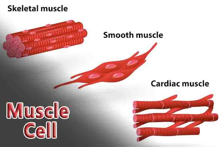
There are three main types of muscle cells in the human body: skeletal muscle cells, cardiac muscle cells, and smooth muscle cells. Skeletal muscle cells are attached to bones and allow for voluntary movement, while cardiac muscle cells make up the walls of the heart and are responsible for pumping blood. Smooth muscle cells are found in the walls of internal organs and control involuntary movements, such as digestion.
Muscle cells contract through a process called the sliding filament theory. This involves the interaction of two types of protein filaments, actin and myosin, which slide past each other to create muscle tension and movement.
During exercise, muscle cells are stimulated to contract and relax repeatedly, which can lead to an increase in muscle size and strength over time. Exercise also triggers the production of new muscle cells through a process called muscle hypertrophy.
Yes, muscle cells have the ability to regenerate to some extent. When muscle cells are damaged, such as through injury or disease, satellite cells (specialized stem cells) can be activated to help repair and regenerate the damaged tissue. However, severe or chronic damage can lead to scarring and loss of muscle function.